

- Information
Examination instruments
-
OCT(Optical Coherence Tomography)
-
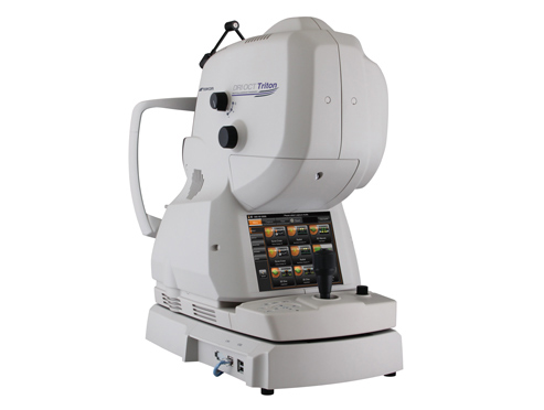
- Take photograph of retina. This test can help to determine detailed data of your eyes like a MRI. This test is used to examine the glaucoma and loss of eye sight.
-
-
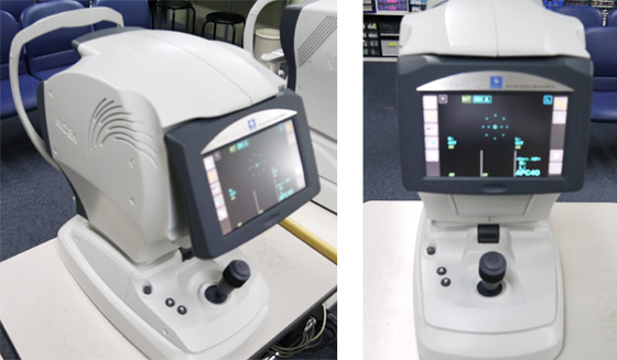
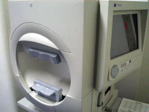
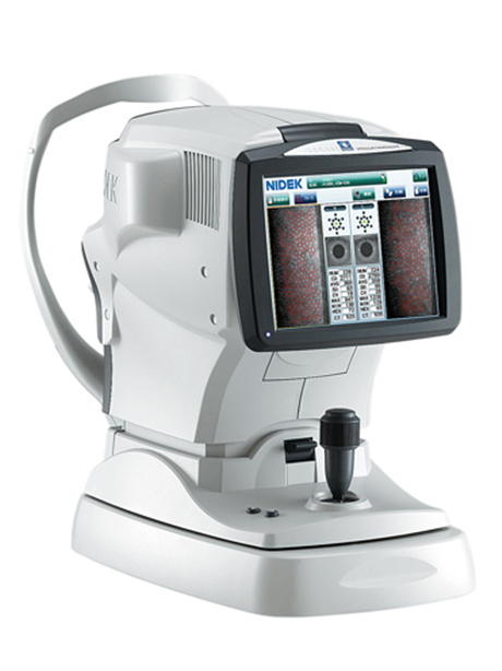
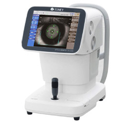
Auto Ref/Kerato/Tono/Pachymeter TONOREF™ III
-
-
Overview; This machine enables to have the refraction test and measure intraocular pressure together.You will be asked to look through the device. We use a non-contact tonometry with an “air puff”to measure your intraocular pressure.
Purpose:Refraction test can help to determine the extent of poor vision and monitor a person who is being treated for an eye disease. Eye pressure measurement is used for determining whether you have glaucoma, a disease which causes pressure to build up inside your eyes and may result in blindness.
Medical device approval number: 227AABZ00095000
-
-
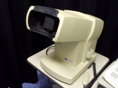
Anterior segment tomography (Pentacam)
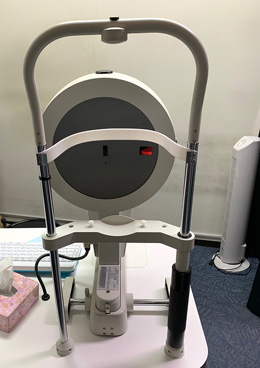
- The test with Pentacam will show the shape of your cornea in only two seconds. This instrument greatly improves the speed and accuracy of the test.
They use Pentacam to check your corneal astigmatism, fitting simulation of contact lenses, and find keratoconus in early stage.
Also, it will show corneal curvature. The image given by Pentacam displays how much your cornea is deformed, how thick it is and the shape of the astigmatism in clear color form.
Concerning cataract and glaucoma, we can measure how much your vision is impaired and how much cataract is developing. Also, we can analyze the shape of the anterior chamber, so it helps us diagnose glaucoma.
-
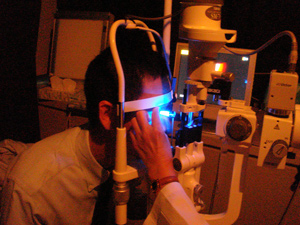
Retinal Examination(Ophthalmoscopy)
-
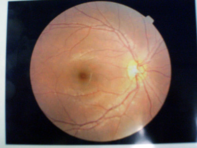
- This exam needs to open your pupil. Eye drops may be placed in your eyes to widen (dilate) your pupils. This test can be help to determine cataract and another inflammation of crystalline lens.
-
-
Pseudoisochromatic plates
-
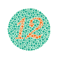
- Ishihara test is one of the most common examination for color blindness. Patients would see a set of dotted plates which show some numbers hidden in the design, and the it is efficient for detecting colour vision deficiency. We also have Panel D-15 test which examines the type and its severity of the colour blindness.
-
-
Humphrey Field Analyzer
- This test can help to determine the loss of your eye sight. You will rest your chin and forehead on a support that keeps your head steady. There are FDT, Humphrey Field Analyzer and Gold man Field Analyzer in our eye clinic.
-
Goldmann Perimetry
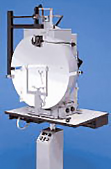
- Goldmann Perimeter examines the range and the sensitivity of your vision. While you are looking at the fixation lamp, an examiner move the light from outside towards the center, and tests the range of your vision.
Unlike Humphrey Field Analyzer and FDT Perimetry, this method is able to test the entire range of peripheral vision, and provides the result as a map which helps confirm a diagnosis of the cause of the symptom.
-
Frequency doubling technology (FDT)
-
- Frequency doubling technology (FDT) is an instrument to determine field vision loss. It is easily used and has high sensitivity to determine the perimeter loss of vision of glaucomatous, retinal, neurological disorder patients.
-
-
Specular Microscope
- Cataract surgery, LASIK and wearing contact lens for a long time may cause loss of corneal endothelial cells. This instrument examines and analyzes the corneal endothelium, and is efficient fo diagnosis of corneal disease too.
-
Amsler Grid Test
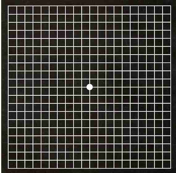
- By taking a look of test chart, we can check distortion and lack of your vision.
-
Allergy test (Blood test)

- It can determine the cause of 39 types of allergies at once.
-
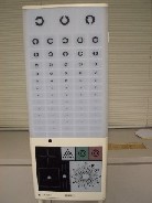
Visual acuity testing
-
Overview: To determine the smallest letters a person can read on a standardized chart. In the test, you will be asked to tell the direction like "up", "down", "right", "left".
Purpose: To find the "best corrected vision" for any visual problems.
-
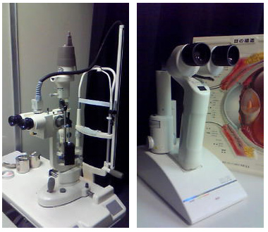
Slit-lamp microscopy
-
Overview: To examine the structures nearby your eyes. In the test, you will be asked to rest your chin and forehead on a support to keep your head steady.
Purpose: The ophthalmologist will examine the cornea, iris, lens and the anterior chamber.
-
Applanation tonometry
-
- This test measures fluid pressure inside your eye. In this test, an equipment called slit lamp will be used, and you will be asked to put your chin and forehead on the support. The doctor will use a flat-tipped cone and put it on your eye. The tonometry measures the amount of pressure needed to flatten your cornea.
-
-
Gonioscopy
- Checks for blockages in the area where fluid drains out of the eye.
This test can help to determine whether or not you need laser exam. Also this exam used to look at the front part of the eye between the cornea and the iris.
-
Flow examination
- We test for obstruction of the lacrimal passage from the punctum to the canaliculi and nasolacrimal duct by running saline through. This test is efficient to diagnose the obstructive illnesses or infections in the lacrimal passage.
-
Optical ocular axial length measuring instrument
- It provides many measurements required for cataract surgery at once.
For example, it can measure the axial length, corneal curvature, anterior chamber depth and lens thickness without touching eyes.
-
Ocular axial/ Corneal thickness measurement instrument (A-mode echogram)
-
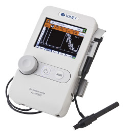
- It is ultrasonic ocular axial and corneal thickness measurement instrument.
These results are measured by using refractive echo and displayed as waveforms.
-
-
Ophthalmoscope
-
-
Overview: To examine the back of your eye using a magnifying instrument (ophthalmoscope) and a light source.
Purpose: The ophthalmologist detects eye diseases such as hypertension, diabetes and atherosclerosis.
-